 Retroviruses & Structural Biochemistry
 Retroviruses & Structural Biochemistry
 are characterized by their ability to gain resistance from therapeutic or vaccinal intervention through the accumulation of escape mutations in the viral genome and/or the interaction of regulatory viral proteins with cellular pathways. Understanding the structural determinants of the functions of the viral proteins is crucial for the development of efficient antiviral therapies.
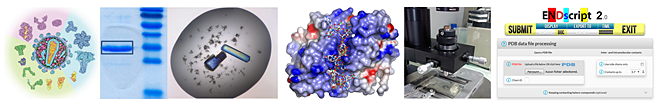 ), observed a new dimeric form of an avian integrase domain (Ballandras et al., 2011 ) and determined the crystal structures of the matrix and capsid proteins of the Feline Immunodeficiency Virus (FIV) (Serrière et al., 2013 ; Folio et al., 2017 ).
 , in coll. with O. Marcillat), the bacterial type 2 secretion system (Pineau et al., 2014 , in coll. with V. Shevchik) and the Ser/Thr kinase StkP of S. pneumoniae (Righino et al., 2017 , in coll. with MC. de Rosa; Zucchini et al., 2018 , in coll. with C. Grangeasse). / ENDscript for protein sequence and structure analyses, which counts more than 15,000 users per year (Robert & Gouet, 2014 ).
 By clicking on the PDB logo, you will be redirected to the Protein Data Bank website presenting the extensive list of the structures we solved @IBCP.
 |

Back to top
| Crystal Structure of the Full-Length Feline Immunodeficiency Virus Capsid Protein |
|---|
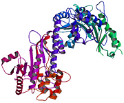
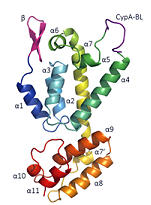 Feline immunodeficiency virus (FIV) is a member of the Retroviridae family. It is the causative agent of an acquired immunodeficiency syndrome (AIDS) in cats and wild felines. Its capsid protein (CA) drives the assembly of the viral particle, which is a critical step in the viral replication cycle. Here, the first atomic structure of full-length FIV CA to 1.7 Å resolution is determined. The crystallized protein exhibits an original tetrameric assembly, composed of dimers which are stabilized by an intermolecular disulfide bridge induced by the crystallogenesis conditions. The FIV CA displays a standard α-helical CA topology with two domains, separated by a linker shorter than other retroviral CAs. The β-hairpin motif at its amino terminal end, which interacts with nucleotides in HIV-1, is unusually long in FIV CA. Interestingly, this functional β-motif is formed in this construct in the absence of the conserved N-terminal proline. The FIV CA exhibits a cis Arg–Pro bond in the CypA-binding loop, which is absent in known structures of lentiviral CAs. This structure represents the first tridimensional structure of a functional, full-length FIV CA.
Folio C, Sierra N, Dujardin M, Alvarez G & Guillon C (2017). Viruses. 9:335
|
| Biophysical characterization and crystal structure of the Feline Immunodeficiency Virus p15 matrix protein |
|---|
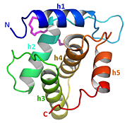 Our biochemical study of the p15 matrix protein of the Feline Immunodeficiency Virus (FIV) revealed that it forms a stable dimer in solution under acidic conditions and at high concentration, unlike other retroviral matrix proteins. We determined the crystal structure of full-length FIV p15 to 2 Å resolution and observed a helical organization of the protein, typical for retroviral matrix proteins. A hydrophobic pocket that could accommodate a myristoyl group was identified, and the C-terminal end of FIV p15, which is mainly unstructured, was visible in electron density maps. As FIV p15 crystallizes in acidic conditions but with one monomer in the asymmetric unit, we searched for the presence of a biological dimer in the crystal. No biological assembly was detected by the PISA server, but the three most buried crystallographic interfaces have interesting features: the first one displays a highly conserved tryptophan acting as a binding platform; the second one is located along a 2-fold symmetry axis and the third one resembles the dimeric interface of EIAV p15. Because the C-terminal end of p15 is involved in two of these three interfaces, we investigated the structure and assembly of a C-terminal-truncated form of p15 lacking 14 residues. The truncated FIV p15 dimerizes in solution at a lower concentration and crystallizes with two molecules in the asymmetric unit. The EIAV-like dimeric interface is the only one to be retained in the new crystal form and could therefore correspond to the one of FIV p15 in solution.
Serrière J, Robert X, Perez M, Gouet P & Guillon C (2013). Retrovirology. 10:64
|
| A crystal structure of the catalytic core domain of an avian sarcoma and leukemia virus integrase suggests an alternate dimeric assembly |
|---|
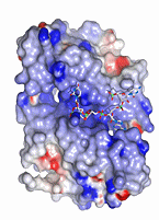 Integrase (IN) is an important therapeutic target in the search for anti-Human Immunodeficiency Virus (HIV) inhibitors. This modular enzyme is hard to crystallize. A first structural result was obtained with the IN catalytic core domain (CCD) of the avian Rous Sarcoma Virus (RSV). A ribonuclease-H like motif was revealed as well as a dimeric interface stabilized by two pairs of α-helices. These structural features have been validated in other structures of IN CCDs. We have determined the crystal structure of the Rous-associated virus type-1 (RAV-1) IN CCD to 1.8 Å resolution. RAV-1 IN shows a standard activity for integration and its CCD differs in sequence from that of RSV by a single accessible residue in position 182 (substitution A182T). Surprisingly, the CCD of RAV-1 IN associates itself with an unexpected dimeric interface characterized by three pairs of α-helices. A182 is not involved in this novel interface, which results from a rigid body rearrangement of the protein at its dimeric surface. A new basic groove that is suitable for single-stranded nucleic acid binding is observed at the surface of the dimer. We have determined the structure of the mutant A182T of RAV-1 IN CCD and obtained a RSV IN CCD-like structure with two pairs of buried α-helices at the interface. Our results suggest that the CCD of avian INs can dimerize in more than one state. Such flexibility can further explain the multifunctionality of the retroviral IN, which beside integration of dsDNA is implicated in different steps of the retroviral cycle in presence of viral ssRNA.
Ballandras A, Moreau K, Robert X, Confort MP, Merceron R, Haser R, Ronfort C & Gouet P (2011). PLoS One. 6:e23032
|
| The substrate-free and -bound crystal structures of the duplicated taurocyamine kinase from the human parasite Schistosoma mansoni |
|---|
 The taurocyamine kinase from the blood fluke Schistosoma mansoni (SmTK) belongs to the phosphagen kinase (PK) family and catalyzes the reversible Mg2+-dependent transfer of a phosphoryl group between ATP and taurocyamine. SmTK is derived from gene duplication, as are all known trematode TKs. Our crystallographic study of SmTK reveals the first atomic structure of both a TK and a PK with a bilobal structure. The two unliganded lobes present a canonical open conformation and interact via their respective C- and N-terminal domains at a helix-mediated interface. This spatial arrangement differs from that observed in true dimeric PKs, in which both N-terminal domains make contact. Our structures of SmTK complexed with taurocyamine or L-arginine compounds explain the mechanism by which an arginine residue of the phosphagen specificity loop is crucial for substrate specificity. A SmTK crystal was soaked with the dead-end transition state analog (TSA) components taurocyamine-NO3--MgADP. One SmTK monomer was observed with two bound TSAs and an asymmetric conformation, with the first lobe semiclosed and the second closed. However, isothermal titration calorimetry and enzyme kinetics experiments showed that the two lobes function independently. A small angle X-ray scattering model of SmTK-TSA in solution with two closed active sites was generated.
Merceron R, Awama AM, Montserret R, Marcillat O & Gouet P (2015). J. Biol. Chem. 290:12951-63
|
| Deciphering key features in protein structures with the new ENDscript server |
|---|
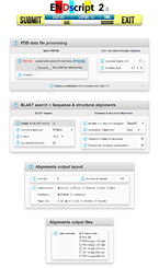 ENDscript 2 is a friendly webserver for extracting and rendering a comprehensive analysis of primary to quaternary protein structure information in an automated way. This major upgrade has been fully re-engineered to enhance speed, accuracy and usability with interactive 3D visualization. It takes advantage of the new version 3 of ESPript, our well-known sequence alignment renderer, improved to handle a large number of data with reduced computation time. From a single PDB entry or file, ENDscript produces high quality figures displaying multiple sequence alignment of proteins homologous to the query, colored according to residue conservation. Furthermore, the experimental secondary structure elements and a detailed set of relevant biophysical and structural data are depicted. All this information and more are now mapped on interactive 3D PyMOL representations. Thanks to its adaptive and rigorous algorithm, beginner to expert users can modify settings to fine-tune ENDscript to their needs. ENDscript has also been upgraded as an open platform for the visualization of multiple biochemical and structural data coming from external biotool webservers, with both 2D and 3D representations. ENDscript 2 and ESPript 3 are freely available at https://endscript.ibcp.fr and https://espript.ibcp.fr .
Robert X & Gouet P (2014). Nucleic Acids Res. 42:W320-4
|
Back to top
| Internal IBCP |
|---|
|
Vincent ChaptalMMSB, CNRS – Université de Lyon Christophe GrangeasseMMSB, CNRS – Université de Lyon Guillaume LaunayMMSB, CNRS – Université de Lyon Juliette MartinMMSB, CNRS – Université de Lyon Cédric OrelleMMSB, CNRS – Université de Lyon Bernard VerrierLBTI, CNRS – Université de Lyon |
| External |
|---|
|
Guzmán Álvarez TouronUniversidad de la República - Paysandú, Uruguay Yannick BlanchardViral Genetics and Biosafety - ANSES Ploufragan, France Laure GuyÉcole Normale Supérieure - Lyon, France Xavier HanoulleNMR & Molecular Interactions - CNRS Villeneuve d'Ascq, France Vincent ParissiFundamental Microbiology and Pathogenicity Lab - CNRS Bordeaux, France Stéphane PaulGroupe sur l'Immunité des Muqueuses et Agents Pathogènes - Faculté de Médecine de Saint-Etienne, France Corinne RonfortPathogenicity and Virus Vaccine Lab - INRA Grenoble, France Maria-Cristina de RosaInstitute of Chemistry of Molecular Recognition - Rome, Italy Guy SchoehnInstitute of Structural Biology - Grenoble, France Vladimir ShevchikMicrobiology, Adaptation and Pathogenesis Lab - INSA Villeurbanne, France |
Back to top
Back to top

|

|

|

|

|

|

|

|

|

|

|
Back to top
 Biochemistry room 1 |
 Biochemistry room 2 |
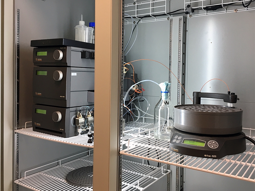 FPLC system 1 |
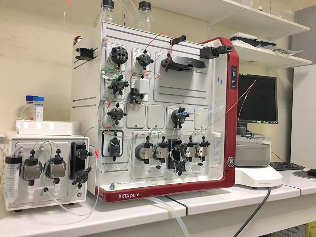 FPLC system 2 |
 DLS system |
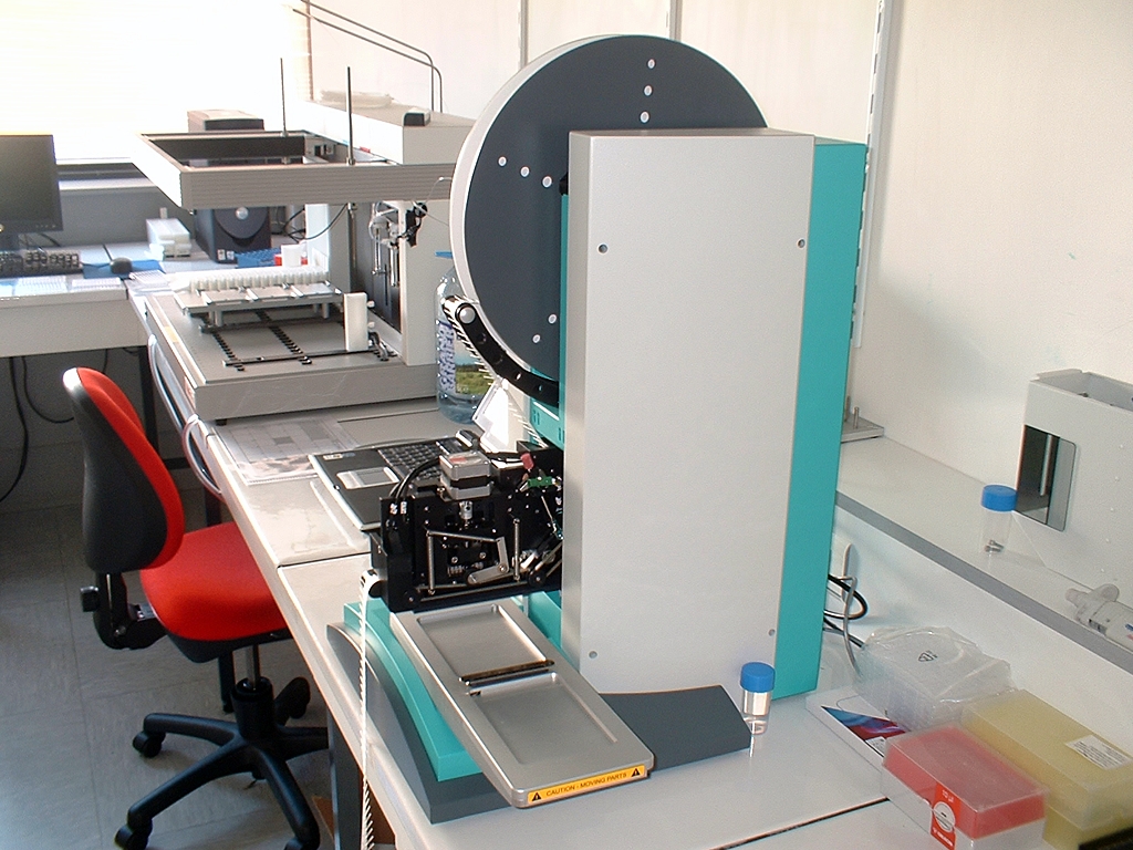 Crystallization robot |
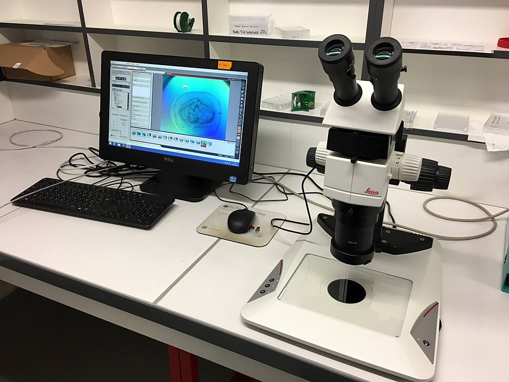 Microscopes room |
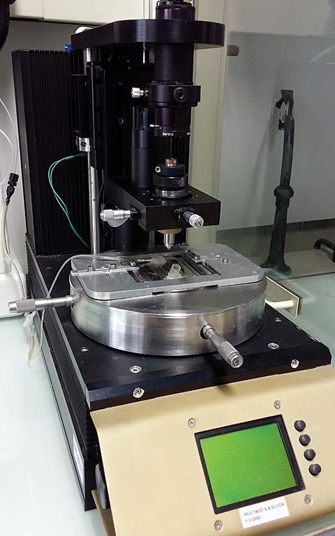 Magnetic tweezers setup |
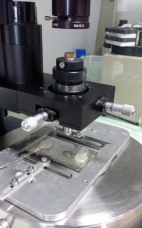 Magnetic tweezers setup |
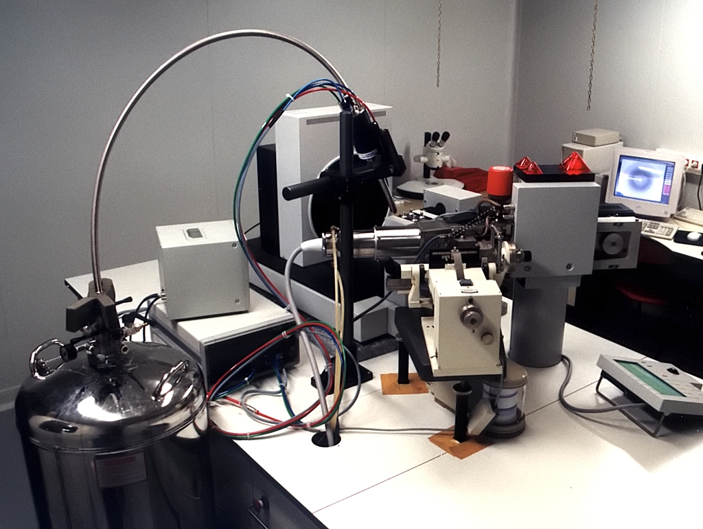 X-ray diffraction setup |
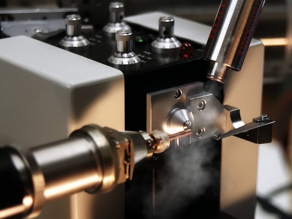 Cryo data collection |
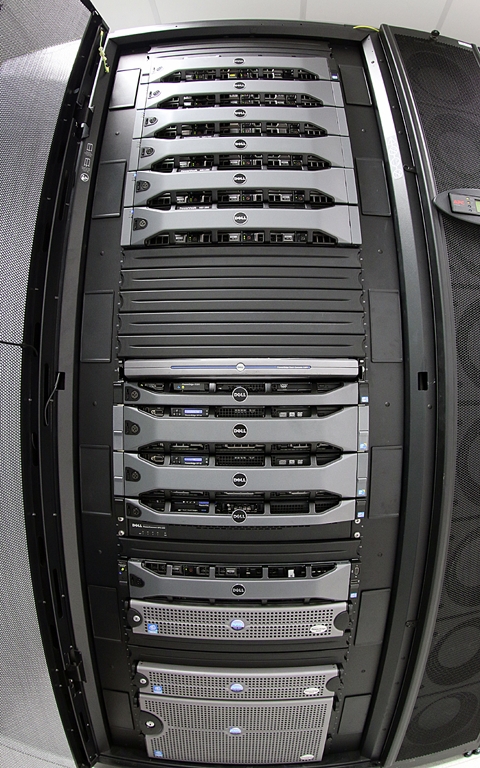 Calculation servers |
Back to top
 Francesca FIORINI CR CNRS |
 Francine GERARD-BARAGGIA MC Univ. Lyon 1 |
 Patrice GOUET PR Univ. Lyon 1 |
 Christophe GUILLON CR CNRS |
 Xavier ROBERT IR CNRS |
Back to top


 Protéines, 7 passage du Vercors,